




Conference presentation
Acute Horner Syndrome identified by Pupillography revealing Internal Carotid Artery Dissection
European Neuro-Ophthalmology Society (EUNOS) Meeting, 2026
doi:
10.1080/01658107.2026.2651025View publication
Study details
Introduction
Acute anisocoria with ipsilateral ptosis, particularly when the pupillary asymmetry is greater in the dark, should raise suspicion for
Horner syndrome and prompt urgent evaluation for potentially life-threatening causes such as internal carotid artery (ICA) dissection. Early recognition in the neuro-ophthalmology setting is critical to prevent cerebral ischemic complications.
Case Report
- A 38-year-old woman presented with a smaller right pupil and mild right ptosis for 5 days, accompanied by right neck pain and facial pressure. She had recently been physically active. Exam showed right miosis (more pronounced in dim light) with briskly reactive pupils and mild ptosis.
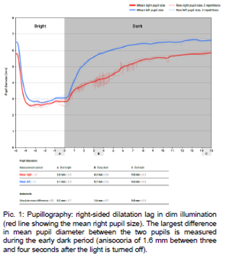
- Pupillography (neos® system) demonstrated a clear right-sided dilatation lag, providing objective confirmation of oculosympathetic dysfunction. Pharmacological testing for Horner syndrome was not pursued because the lopidine test could not be realiably interpreted in cases of recent onset.
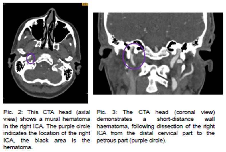
- Contrast-enhanced computed tomography (CT) of the head and CT angiography (CTA) revealed a short-segment mural hematoma consistent with right ICA dissection, extending from the distal cervical segment to the transition into the petrous segment. No intracranial hemorrhage or cerebral ischemia was detected.
Management
- The patient was admitted for monitoring and started on antiplatelet therapy with Aspirin 100mg.
- Cervical artery duplex ultrasound showed no significant stenosis
- With no signs of connective tissue disorder and a history of intense physical activity, the dissection was considered likely provoked.
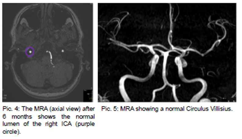
- MR angiography (MRA) at 6-month follow-up showed normalization of the vessel lumen.
Discussion
- Acute Horner syndrome with ipsilateral neck/facial pain is a classic red flag for ICA dissection.
- As pupillary findings may be subtle, objective testing (e.g., pupillography) can support clinical suspicion, expedite emergency imaging, and occasionally a pharmacologic testing is not necessary any more due to the characteristic appearance with dilation lag in pupillography.
- Prompt vascular imaging with CTA or MRA is essential for diagnosis.
- Early antithrombotic therapy (antiplatelet or anticoagulaton, depending on clinical context) reduces thromboembolic and ischemic risk.
- Painful acute Horner syndrome should be treated as an emergency until ICA dissection is excluded.


